Navigation
Mammography
Mammography is a specialised imaging procedure that uses low-dose X-Rays to capture detailed images of breast tissue. This essential diagnostic tool plays a vital role in detecting and diagnosing breast conditions, including breast cancer, at an early stage. By identifying tissue changes, such as small lumps or calcifications, mammograms can detect abnormalities that might not be noticeable during a physical examination, supporting proactive and preventative healthcare.
FAQs
Yes, mammograms require an appointment. You can schedule your exam by contacting our team via phone, visiting in person, or submitting your referral. Please have details about prior mammograms and family history available, as this information may help determine your eligibility for Medicare rebates.
Preparation instructions will be provided during booking. We recommend wearing a two-piece outfit for convenience and avoiding the use of deodorant, talcum powder, or perfume on the day of your exam, as these substances can interfere with image quality. Arrive at least 15 minutes early to complete any necessary paperwork.
Bring the following items:
- Your referral form.
- Any previous imaging reports or mammograms.
- Your Medicare or DVA card, if applicable.
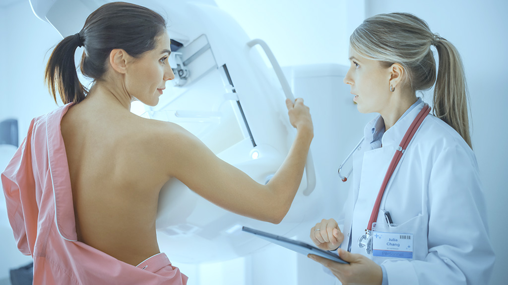
A staff member will guide you to the examination room, explain the process, and help you prepare. You may be asked to wear a gown. During the mammogram, your breast tissue will be positioned on a flat plate and gently compressed with a paddle. This compression spreads the tissue for clearer imaging and minimises the radiation required. While some brief discomfort may occur, it is typically limited to the duration of the image capture. After the procedure, a specialist radiologist will analyse the images and prepare a report for your referring doctor.
Costs depend on eligibility for a Medicare rebate, which may be influenced by factors such as family history or symptoms. Diagnostic mammograms are generally eligible for a rebate, while screening mammograms may involve an out-of-pocket expense without rebate coverage.
Reports are typically available within 24 to 48 hours. If your doctor has requested immediate results, please notify our staff. Urgent cases are prioritised, and our team will work with the radiologist to expedite your report if necessary.

